Virtual Blood Smear Lab
Lab Objectives:
* Identify the component cells of a typical blood smear.
* Compare and contrast the compound light microscope and electron microscope views of blood cells.
* Distinguish the different classes of white blood cells and the conditions under which each would be expected to dominate.
Introduction:
Blood provides a mechanism by which nutrients, gases, and wastes can be transported throughout the body. It consists of a number of cells suspended in a fluid medium known as plasma. The cells of the blood consist of erythrocytes, platelets, and leukocytes or white blood cells. Erythrocytes are responsible for transporting gases,
The cells of the blood are important because they are a readily accessible population whose morphology, biochemistry, and ecology may give indications of a patient's general state or clues to the diagnosis of disease. Physicians may call for a "complete blood count" or CBC which measures the cells that are in your blood. Collecting a CBC is quite simple, just as long as you are comfortable with a little needle. The Phlebotomist or examiner will collect some blood from your arm send it off to the local lab for further testing. You are then free to leave and go about your day as usual. If more information is required a differential white cell count may be done. A differential white cell count identifies the types of white blood cells present and in the percentages present. For this reason, the complete blood count (CBC) and the differential white cell count, are routinely used in clinical medicine. It is very important to be able to recognize normal blood cells and to distinguish pathological cells from the normal variants.
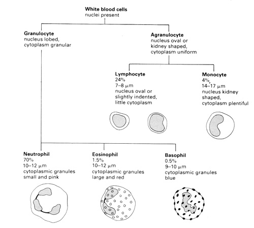
The identification of blood cells is based primarily on observations of the presence or absence of a nucleus and cytoplasmic granules. Other helpful features are cell size, nuclear size and shape, chromatin appearance, and cytoplasmic staining. The chart below explains what to look for in the effort to identify the component cells of a blood smear.

Career Connections:
What is a Phlebotomist? Click HERE to learn what a phlebotomist does and how to become one.
Sometimes blood samples are drawn to learn what has been ingested or taken intravenously by someone who has passed away. Forensic investigators can piece together what happened by sometimes collecting samples of blood that are taken during the autopsy. These samples are then sent to "toxicology". Click HERE to learn more about what a toxicologist might do.
Blood stains and residue can also help investigators learn more about their cases. Click HERE to learn more about Bloodstain Pattern Analysis. The popular TV series "Dexter" helped to bring this obscure career to the forefront of forensics.
Erythrocytes
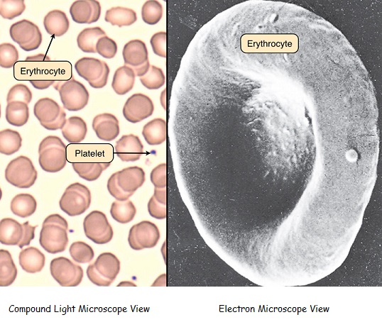
Erythrocytes, or red blood cells, are by far the predominant cell type in the blood smear. They appear as biconcave discs of uniform shape and size (7.2 microns) that lack organelles and granules. Red blood cells are "anuclear" cells which means they do not have a nucleus. Therefore red blood cells must be constantly replenished by the body. This process is referred to as hematopoiesis and takes place in the bone marrow of long bones. Red blood cells have a characteristic pink appearance due to their high content of hemoglobin. The central pale area of each red blood cell is due to the concavity of the disc. Also visible in this slide are several platelets, which play a crucial role in the blood clotting cascade.

Neutrophils
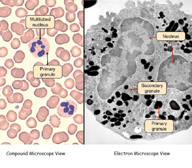
Neutrophils are by far the most numerous of the leukocytes. They are characterized by a nucleus that is segmented into three to five lobes that are joined by slender strands. The cytoplasm of neutrophils stains a pale pink. Its primary (larger) granules contain acid hydrolases and cationic proteins, and its secondary (smaller) granules contain a variety of antimicrobial substances used to destroy bacteria that they phagocytose during the acute inflammatory response.

Eosinophils
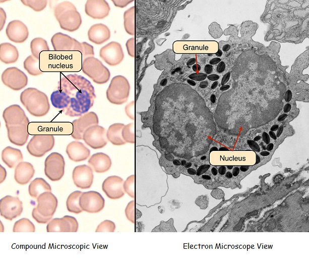
Eosinophils are larger than neutrophils and are distinguished by bilobed nucleus and large red or orange granules of uniform size. These granules contain major basic protein, which is released to kill organisms too large to phagocytose, such as parasites and helminthes (worms). Eosinophils make up between 1 and 3% of the total white blood cells in the human blood.

Basophils
Basophils are intermediate in size between neutrophils and eosinophils and have simple or bilobed nuclei. They contain many coarse purple granules that can vary in size or shape. These granules contain histamine, which is released to cause a vasoactive response in hypersensitivy reactions, and heparin, which is an anticoagulant. Basophils are not phagocytic.

Lymphocytes
Lymphocytes can appear either small or large. The small lymphocyte is about the same size as an erythrocyte and contains a dark nucleus with a thin rim of surrounding cytoplasm. Lymphocytes do not contain visible granules. Small lymphocytes are inactive. Large lymphocytes (10 - 15 microns) contain more cytoplasm than small lymphocytes, and the cytoplasm remains basophilic. Large lymphocytes are active B or T cells. It is not possible to distinguish B- and T-lymphocytes at this level of magnification.

Monocytes
Monocytes are larger than lymphocytes and granulocytes and contain nuclei that often contain an indentation on one side. The cytoplasm contains small granules with lysosomal enzymes and peroxidase. Monocytes are phagocytic cells that are important in the inflammatory response.
